New Surgical Technology First Tested In Humans At Cedars-Sinai Is Giving Fido A Second Chance, Too
Some of man's best friends are wagging their tails - literally -- thanks to human research on a new type of surgical imaging device being pioneered at Cedars-Sinai Medical Center. Nine dogs that would have died of canine Cushing's disease are alive and barking today, and even one cat has been given a new lease on one of its nine lives.
Neurosurgeon Adam N. Mamelak, M.D., had been studying the use of a scope called a VITOM™ for human surgery when he was approached by a group of veterinary endocrinologists and surgeons at VCA West Los Angeles Animal Hospital who were interested in having him teach them to perform similar surgeries in dogs. Some pituitary tumors are extremely common in dogs, and often fatal.
After studying the problem, Mamelak, an expert in minimally invasive pituitary surgery in the Department of Neurosurgery and co-director of the Pituitary Center at Cedars-Sinai, noted that the VITOM device happens to be a nearly perfect fit for use in dogs with pituitary tumors. He agreed to proctor the animal hospital veterinarians in performing potentially life-saving canine neurosurgery to remove these tumors.
This arrangement benefits both canine and human patients because after a tumor is removed, Mamelak takes tumor tissue back to Cedars-Sinai's laboratories for study, and research teams have already begun to make important observations about treating the tumors with certain drugs.
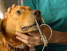
Only one other group in the world - in the Netherlands - is known to be regularly attempting a similar procedure in dogs. Both groups use what is called a transsphenoidal approach, creating a tiny hole in the back of the mouth to enter the skull at the base of the brain and remove the tumor. But because dogs have long snouts, Mamelak says there isn't much to see. The VITOM, which is also called an exoscope, solves this problem by providing up to 12 times magnification and projecting the operating field onto a large high-definition video monitor. This gives the surgeon a vastly larger and sharper view of the tumor and the surrounding brain structures, making removal safer and easier.
Mamelak says veterinary medicine is becoming increasingly sophisticated and is almost as technologically advanced as human medicine. The exoscope is cutting-edge human surgery technology that has made an early jump - at least in a limited way - to a veterinary application.
"I've been training the veterinarians to use the exoscope," Mamelak says. "They've never done any significant neurosurgery, let alone through a tiny hole, but they're getting better and better. By adopting technology developed for humans - and tested initially here at Cedars-Sinai - to veterinary medicine, we are able to provide a technological leap that makes the procedure more accessible to veterinarians and their patients."
So far, the operation has been performed on 14 animals. Eight dogs and one cat have survived and are doing very well, according to Mamelak.
Although canine pituitary tumors are not identical to those in humans, they are very similar, making the canine disease a very good model to study for understanding human illness as well. Interestingly, the most common tumors found in dogs - those that produce too much of a hormone called ACTH and cause Cushing's disease - are extremely rare in humans. Cushing's disease occurs in only about one in every 1 million people, but there are more than 100,000 cases in dogs each year in the U.S. alone.
Symptoms include increased thirst and urination, diabetes, hair loss, thinning skin, increased appetite, and abdominal enlargement. Without treatment, the canine disease is fatal, and the few existing drugs for the condition are usually not curative, have serious side effects, and can be very expensive.
All of the veterinary work is done with the expressed consent and approval of the pet owners, using strict federal guidelines for humane animal care.
"This research collaboration benefits both humans and canines with these tumors. In addition to saving dogs' lives, it provides a mechanism for early testing of drug therapies that may be useful for humans as well. As we progress with our laboratory studies we are identifying drugs that may treat the tumors. We then hope to be able to give medicines to dogs to shrink their tumors, then monitor the dogs, perform the surgery, and restudy the tissue to see how it was affected by the medicine," says Mamelak, a dog-lover who has a 6-year-old mutt named Maya at home. "This working model really benefits dogs and veterinary medicine as much as it benefits people."
Note: Additional background on Dr. Mamelak and human pituitary surgery:
Although the VITOM exoscope appears to be an excellent tool for several kinds of human surgery and canine pituitary surgery, it is not ideal for human pituitary surgery. For that, Mamelak uses an endoscope - a narrow tube with an HD camera lens at the tip. It is inserted through the nose and the back of the nasal cavity. A small burr hole through bone allows the endoscope and operating instruments to be positioned directly in the area of the pituitary gland, and the surgical site is displayed on a large HD monitor.
"There are no external cuts, bruises or tissues that need to heal, and because the endoscope provides a wide field of view, we can remove tumors deep inside the brain and we can be sure to get every little last bit of the tumor out," Mamelak says.
Most neurosurgeons who treat pituitary tumors still use an operating microscope, which also provides HD viewing, but the equipment is large and bulky and has a fixed focal point. The deeper the tumor, the narrower the field of vision becomes - almost like tunnel vision. Mamelak trains residents in Cedars-Sinai's neurosurgical residency program in the use of the endoscope because, while the microscope is easier for the surgeon to use, he believes the endoscope provides a much better experience for the patient.
The exoscope is something of a hybrid of the endoscope and the operating microscope. It provides image quality rivaling the microscope but is lightweight, portable and far less costly.
Source:
Cedars-Sinai Medical Center
Wednesday, October 20, 2010
Subscribe to:
Post Comments (Atom)


3 comments:
thanks for the information..friend..quite useful:)
Student Accommodation
That's what were here for...Thanks..
Fantastic. I love when such advances are made! Cushing's is a nasty condition.
Post a Comment